Multilevel Vertebrogenic Pain Successfully Treated with Basivertebral Nerve Ablation
Clinical Question
Can careful clinical evaluation identify multilevel vertebrogenic pain in a patient with persistent chronic axial low back pain despite successful treatment of a separate neuropathic pain generator?
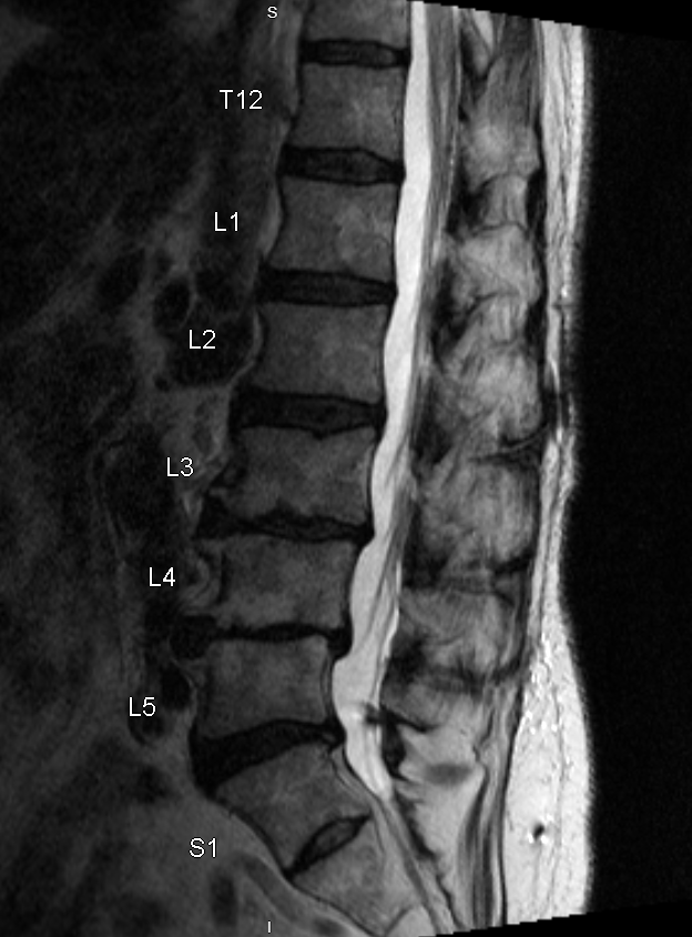 Show labels Enlarge
Sagittal lumbar MRI. Multilevel Modic endplate changes — most advanced at L4–L5, with significant change involving the superior S1 endplate and more subtle changes extending to L3, L2, and L1 — interpreted together with the clinical picture to support multilevel vertebrogenic pain. De-identified · no PHI.
- Treated vertebral levels
- L1 · L2 · L3 · L4 · L5 · S1
- #Imaging findings
- 1Most advanced Modic endplate changeL4–L5 motion segment
- 2Significant Modic endplate changeSuperior S1 endplate
- 3More subtle Modic endplate changesL3, L2, and L1 levels
Reading the Imaging
The MRI is interpreted together with the patient's history, examination, and treatment response.
- Patient Overview
A 76-year-old woman presented with persistent chronic axial low back pain that continued to significantly limit her daily activities and quality of life despite prolonged conservative treatment. Previous treatment included formal physical therapy, activity modification, oral medications — including analgesics, opioids, and neuropathic agents — and interventional pain procedures, as well as Abbott dorsal root ganglion (DRG) stimulation for bilateral lower extremity neuropathic pain.
Although the patient's neuropathic lower extremity symptoms had responded appropriately to DRG stimulation, disabling axial low back pain persisted and continued to interfere with routine daily activities. Among the activities most affected were prolonged sitting, caring for her companion cats, and gardening — activities that had become increasingly difficult because of persistent vertebrogenic pain.
- Clinical Evaluation
Following comprehensive review of the patient's history, physical examination, characteristic pain pattern, previous treatment response, and lumbar MRI, vertebrogenic pain was considered a significant contributor to the patient's persistent axial low back pain.
Lumbar MRI demonstrated multilevel Modic endplate changes of varying severity throughout the lumbar spine. The most advanced Modic endplate changes involved the L4–L5 motion segment, with additional significant Modic changes involving the superior S1 endplate and more subtle Modic endplate changes extending to L3, L2, and L1.
When interpreted in conjunction with the patient's history, physical examination, characteristic pain pattern, and unsuccessful response to prolonged conservative treatment, these findings supported multilevel vertebrogenic pain involving the L1 through S1 vertebral levels. A multilevel Basivertebral Nerve Ablation was subsequently performed at L1, L2, L3, L4, L5, and S1 utilizing the Stryker OptaBlate® system.
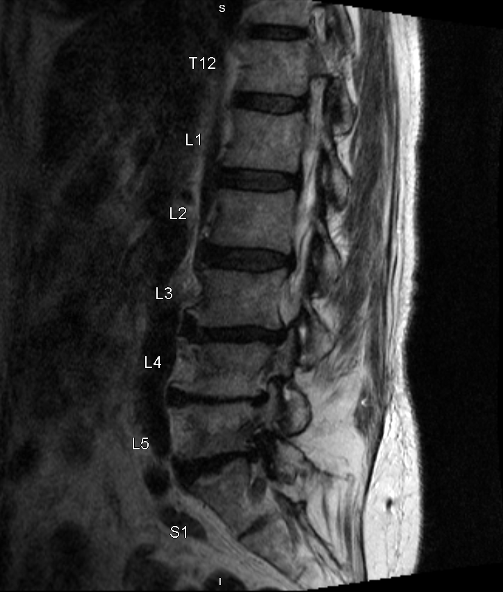
Fluoroscopic Images of Radiofrequency Ablation Probe Placement
Under fluoroscopic guidance, paired anteroposterior (AP) and lateral images were obtained to confirm radiofrequency ablation (heater) probe position within the vertebral body at each treated level — S1 through L1 — using the Stryker OptaBlate® system. The images are organized by region, from the lower lumbar / sacral levels upward to the upper lumbar levels.
Lower Lumbar / Sacral Levels

Anteroposterior (AP) view
L5 and S1 vertebral bodies
Anteroposterior (AP) image demonstrating fluoroscopic localization and final positioning of the Stryker OptaBlate heater probes within the L5 and S1 vertebral bodies.

Lateral view
L5 and S1 vertebral bodies
Lateral image of the same L5 and S1 levels, confirming the depth of the Stryker OptaBlate heater probes within the vertebral bodies.
Mid Lumbar Levels

Anteroposterior (AP) view
L3 and L4 vertebral bodies
Anteroposterior (AP) image demonstrating fluoroscopic localization and final positioning of the Stryker OptaBlate heater probes within the L3 and L4 vertebral bodies.

Lateral view
L3 and L4 vertebral bodies
Lateral image of the same L3 and L4 levels, confirming the depth of the Stryker OptaBlate heater probes within the vertebral bodies.
Upper Lumbar Levels

Anteroposterior (AP) view
L1 and L2 vertebral bodies
Anteroposterior (AP) image demonstrating fluoroscopic localization and final positioning of the Stryker OptaBlate heater probes within the L1 and L2 vertebral bodies.

Lateral view
L1 and L2 vertebral bodies
Lateral image of the same L1 and L2 levels, confirming the depth of the Stryker OptaBlate heater probes within the vertebral bodies.
The Clinical Course
Improvement following BVNA is frequently progressive; early symptoms are not the final outcome.
- Early Recovery
At the one-week postoperative follow-up, the patient reported approximately 20% improvement in previously described axial low back pain. Postoperative healing was appropriate, and no procedure-related complications were identified.
The patient was advised that recovery following Basivertebral Nerve Ablation is often progressive and that continued improvement is expected during the postoperative healing period.
- Progressive Recovery
At the four-week postoperative follow-up, the patient reported approximately 60% improvement in axial low back pain, with steadily improving activity tolerance and daily function.
- Long-Term Outcome
By approximately 12 weeks following the procedure, the patient reported an estimated 75% reduction in the chronic axial low back pain that had previously limited her quality of life.
She described meaningful improvement in her ability to tolerate prolonged sitting, care for her companion cats, and return to light gardening and planting flowers — activities that had become increasingly difficult because of persistent vertebrogenic pain. Overall activity tolerance, independence, and quality of life were substantially improved compared with her pre-procedure baseline.
At a Glance
≈75%
reduction in chronic axial low back pain by ~12 weeks
Patient
76-year-old female
Diagnosis
Persistent chronic vertebrogenic low back pain
Prior treatment
Successful Abbott DRG stimulation for bilateral lower extremity neuropathic pain
Procedure
Six-level BVNA (L1–S1), Stryker OptaBlate® system
One / four weeks
≈20% → ≈60% improvement
Twelve weeks
≈75% improvement · returned to sitting, cats, and gardening
Clinical Pearl
Patients with chronic spinal pain frequently have more than one pain generator. Successful treatment of neuropathic lower extremity pain with dorsal root ganglion stimulation does not eliminate vertebrogenic pain arising from the vertebral endplates. Careful clinical evaluation remains essential to identify persistent pain generators and match each with the most appropriate treatment. Ultimately, successful treatment of vertebrogenic pain is measured not only by pain reduction, but by meaningful improvement in function, independence, and the ability to return to activities that improve quality of life.
Continue Your Learning
Continue exploring the educational library at your own pace.
Have similar symptoms?
Every case is individual. A personal evaluation can help determine whether an assessment for vertebrogenic pain may be appropriate for you.