Understanding Modic Changes
Modic changes are MRI findings involving the vertebral endplates and adjacent bone marrow. In some patients they may be associated with vertebrogenic pain, but they are only one part of a complete clinical evaluation.
What You'll Learn
Approx. 6–8 min read- What a vertebral endplate is
- What Modic changes look like on an MRI
- What they may mean for low back pain
- How they connect to vertebrogenic pain and BVNA
What Is an Endplate?
Most patients have never heard the term. An endplate is the thin surface of bone at the top and bottom of each vertebral body — the part that faces the disc.
- The disc sits between two vertebrae and cushions them.
- The endplate is the bone surface where the vertebra meets the disc.
- The vertebral body is the main, block-shaped part of the bone.
How Do Modic Changes Develop?
The discs act like shock absorbers. As they lose height and cushioning over time, greater stress may be transferred to the endplates — where MRI can later show changes.
- Step 1 of 4: A cushioned segment. The discs act like shock absorbers, cushioning the vertebrae and protecting the endplates.
- Step 2 of 4: Less cushioning. As discs lose height and cushioning over time, the way load is shared across the segment can change.
- Step 3 of 4: More stress on the endplates. With less cushioning, greater stress may be transferred to the vertebral endplates.
- Step 4 of 4: Changes that MRI may show. Over time, an MRI may show changes in these endplate areas, which radiologists call Modic changes.
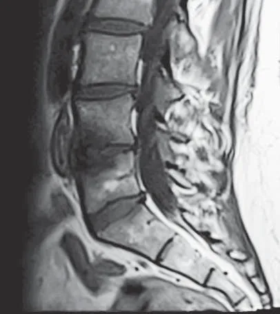
Modic Type 1
Modic Type 1 changes are one pattern radiologists may describe near the vertebral endplates. In general terms, they are often thought to reflect a more active-appearing change in the bone next to the disc.
- A pattern seen near the endplates on MRI.
- Often described as a more 'active' appearance.
- One finding among many that a clinician considers.
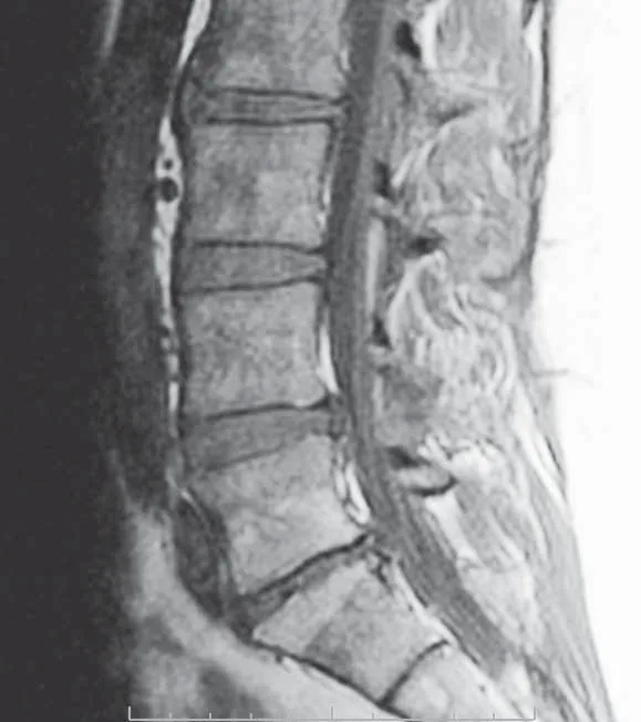
Modic Type 2
Modic Type 2 changes are another pattern near the endplates. In general terms, they are often described as a more 'settled' appearance in the adjacent bone.
- Also seen near the endplates on MRI.
- Often described as a more 'settled' appearance.
- Like Type 1, it is interpreted alongside the full clinical picture.
Type 1 and Type 2 on MRI
MRI scans are read in two common “weightings” — called T1 and T2 — that make tissues look lighter or darker in different ways. Radiologists describe Modic changes by the pattern they show across both. Neither type is “better” or “worse”; they are simply different appearances a clinician interprets in context.
Modic Type 1
Often a more “active” appearance
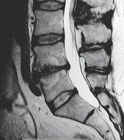
On a T1-weighted image the endplate change tends to look darker; on a T2-weighted image it tends to look brighter.
Modic Type 2
Often a more “settled” appearance
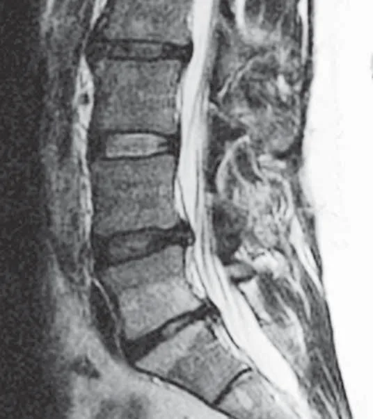
On both T1- and T2-weighted images the endplate change tends to look brighter.
De-identified MRI examples © Boston Scientific — physician-authorized educational use.
Do Modic Changes Always Cause Pain?
No — not always.
Many imaging findings can be present with or without symptoms. Some people have Modic changes and little or no pain; others have pain without these findings. Imaging and symptoms do not always line up.
That is why physicians interpret MRI findings together with your history and physical examination — never from an image alone.
How Physicians Use MRI Findings
Modic changes are one piece of the puzzle. A physician weighs several things together — never one finding alone.
History
How your pain started, how long it has lasted, and how it behaves.
Physical examination
What a hands-on examination shows about your spine and movement.
Pain pattern
Where the pain is, and what makes it better or worse.
Response to prior treatment
How your pain has responded to previous, conservative care.
Imaging
What the MRI shows, including any Modic changes near the endplates.
All together
These are weighed as a whole — not from a single finding.
How Do Modic Changes Relate to Vertebrogenic Pain?
In some appropriately selected patients, Modic changes may support the diagnosis of vertebrogenic pain when considered alongside the patient's symptoms, examination, and imaging. They do not, on their own, mean a person has vertebrogenic pain.
How Does This Relate to BVNA?
When a careful evaluation suggests vertebrogenic pain is a meaningful contributor, one option that may be discussed is Basivertebral Nerve Ablation (BVNA). Modic changes are only one part of that picture — they do not, by themselves, mean BVNA is appropriate.
Frequently Asked Questions
Educational answers to a few common questions about Modic changes.
Are Modic changes the same as arthritis?
Do Modic changes always hurt?
Can Modic changes improve or change over time?
Does everyone with Modic changes need treatment?
Can an MRI finding alone diagnose the cause of my pain?
Continue Your Learning
Keep exploring the educational library at your own pace.