Persistent Vertebrogenic Pain Following Prior Lumbar Fusion
Clinical Question
Can persistent vertebrogenic pain remain after an otherwise successful lumbar fusion, and can careful evaluation identify patients who may benefit from Basivertebral Nerve Ablation despite prior spinal instrumentation?
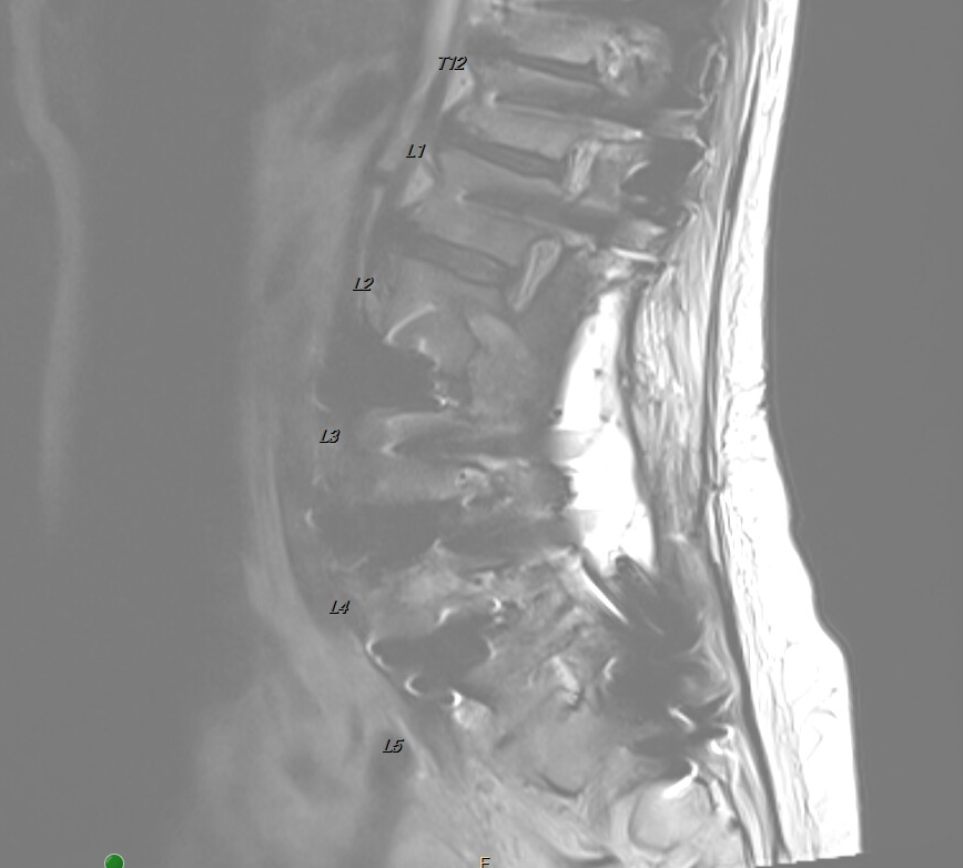 Show labels Enlarge
Sagittal lumbar MRI in a patient with a prior multilevel lumbar fusion. The posterior instrumentation and the persistent vertebral endplate changes adjacent to the previously fused motion segments are visible, interpreted together with the clinical picture to support persistent vertebrogenic pain. De-identified · no PHI.
- Treated vertebral levels
- T12 · L1 · L2 · L3 · L4 · L5 · S1
- #Imaging findings
- 1Persistent vertebral endplate changesAdjacent to the previously fused segments
- 2Prior lumbar fusion instrumentationPosterior pedicle screws and rods
Reading the Imaging
The MRI is interpreted together with the patient's history, examination, and treatment response.
- Patient Overview
A 73-year-old woman presented with persistent, debilitating chronic axial low back pain following a prior multilevel lumbar fusion. Her previous surgery had successfully restored spinal stability and significantly improved her lower extremity symptoms; despite this, she continued to experience severe midline low back pain that progressively limited walking, standing, travel, and the activities she had anticipated enjoying during retirement.
She had undergone extensive conservative treatment, including formal physical therapy, a home exercise program, activity modification, oral medications, opioid therapy, epidural steroid injections, and multiple interventional spine procedures. Despite these treatments, her chronic axial low back pain continued to significantly impair her daily function and quality of life.
- Clinical Evaluation
Comprehensive review of the patient's history, physical examination, prior operative history, and lumbar MRI demonstrated persistent vertebral endplate changes adjacent to the previously fused lumbar motion segments. Although the fusion had successfully addressed spinal instability, the history, physical examination, characteristic pain pattern, and MRI findings remained consistent with persistent vertebrogenic pain arising from damaged vertebral endplates.
The diagnosis was made by correlating the patient's history, physical examination, MRI findings, previous surgical history, and failure of prolonged conservative management — rather than relying upon MRI findings alone.
Because the existing spinal instrumentation did not prevent safe fluoroscopic access to the targeted vertebral bodies, Basivertebral Nerve Ablation was recommended and subsequently performed around the existing instrumentation utilizing the Stryker OptaBlate® system.
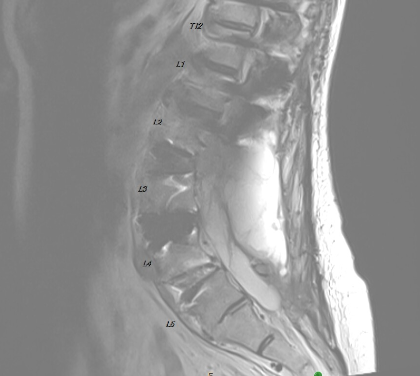
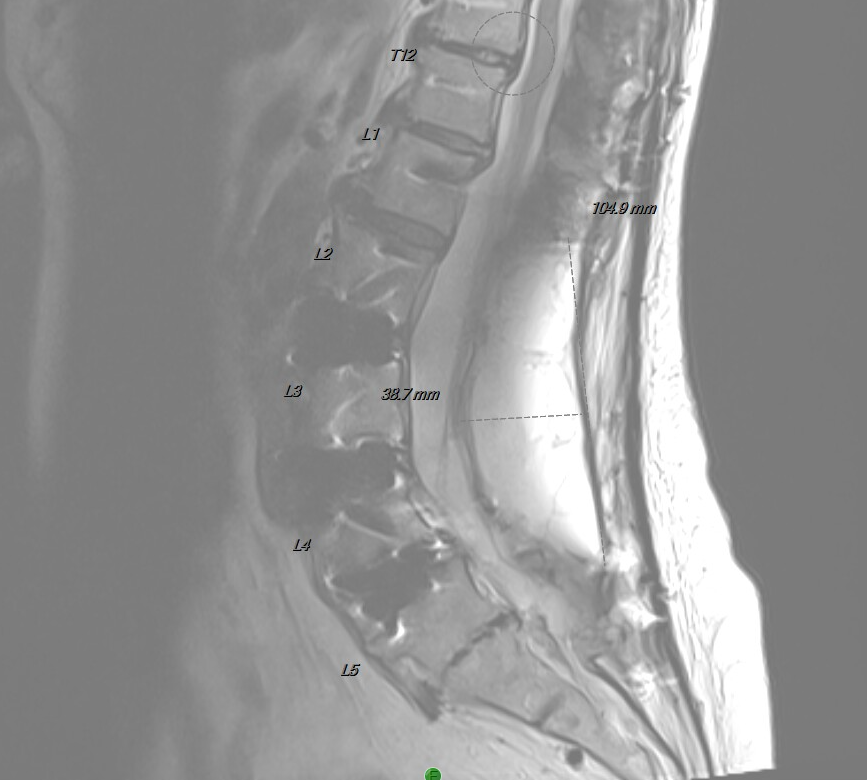
Previous Lumbar Fusion
Before this treatment, the patient had undergone a multilevel instrumented lumbar fusion that successfully restored spinal stability and improved her lower extremity symptoms.

Fluoroscopic Images of Radiofrequency Ablation Probe Placement
Careful fluoroscopic planning allowed safe transpedicular access to the targeted vertebral bodies despite the presence of pedicle screws, rods, and interbody fusion cages. Paired anteroposterior (AP) and lateral images confirmed access and final radiofrequency ablation (heater) probe position at each level — treating from the sacrum upward through the low thoracic spine — using the Stryker OptaBlate® system.

Lateral view
S1 — access channel
Lateral image showing the J-stylette establishing the access channel within the S1 vertebral body, navigated safely alongside the existing instrumentation.

Anteroposterior (AP) view
S1 vertebral body
Anteroposterior (AP) image confirming safe transpedicular access and heater probe position within the S1 vertebral body despite the existing instrumentation.

Lateral view
S1 vertebral body
Lateral image of the S1 heater probe, with the access channel also established at the L5 level.

Anteroposterior (AP) view
S1 and L5 vertebral bodies
Anteroposterior (AP) image demonstrating heater probes positioned within the S1 and L5 vertebral bodies, navigated around the pedicle screws and rods.

Lateral view
S1 and L5 vertebral bodies
Lateral image confirming the depth of the S1 and L5 heater probes within the vertebral bodies.

Lateral view
L5 and L4 vertebral bodies
Lateral image confirming the depth of the L5 and L4 heater probes within the vertebral bodies.

Anteroposterior (AP) view
L3 vertebral body
Anteroposterior (AP) image demonstrating the heater probe within the L3 vertebral body, navigated around the interbody and posterior instrumentation.

Lateral view
L3 vertebral body
Lateral image confirming the depth of the L3 heater probe within the vertebral body.

Anteroposterior (AP) view
L2 vertebral body
Anteroposterior (AP) image demonstrating the heater probe within the L2 vertebral body alongside the instrumentation.

Lateral view
L2 vertebral body
Lateral image confirming the depth of the L2 heater probe within the vertebral body.

Anteroposterior (AP) view
L1 and T12 vertebral bodies
Anteroposterior (AP) image demonstrating heater probes within the L1 and T12 vertebral bodies at the top of the construct.

Anteroposterior (AP) view
T12 and L2 vertebral bodies
Anteroposterior (AP) image demonstrating the Stryker OptaBlate heater probes within the upper lumbar vertebral bodies, from T12 through L2, alongside the existing instrumentation.

Lateral view
T12 and L1 vertebral bodies
Lateral image confirming the depth of the T12 and L1 heater probes within the vertebral bodies.

Lateral view
T12 and L2 vertebral bodies
Lateral image confirming the depth of the Stryker OptaBlate heater probes within the T12 and L2 vertebral bodies.
The Clinical Course
Improvement following BVNA is frequently progressive; early symptoms are not the final outcome.
- Early Recovery
At the one-week postoperative evaluation, the patient reported approximately 10–20% improvement in her characteristic vertebrogenic low back pain while continuing to recover from expected postoperative discomfort. No procedure-related complications were identified.
The patient was counseled that improvement commonly continues during the weeks following Basivertebral Nerve Ablation.
- Progressive Recovery
At the four-week postoperative evaluation, the patient reported approximately 40% improvement in her chronic axial low back pain. Standing tolerance, walking endurance, and overall daily function had noticeably improved.
- Long-Term Outcome
At approximately eight weeks following the procedure, the patient reported approximately 60% overall improvement in the chronic vertebrogenic low back pain that had persisted despite her prior lumbar fusion. She described meaningful improvements in walking, standing, household activities, and participation in normal daily life.
Most importantly, she was once again able to enjoy an active retirement with her husband — including traveling and taking cruises — activities that had become increasingly limited by her persistent back pain before treatment. She reported substantial improvement in both activity tolerance and overall quality of life.
Stabilization and Vertebrogenic Pain Can Coexist
- Two Separate Problems
An important educational message of this case is that successful spinal stabilization and persistent vertebrogenic pain are not mutually exclusive. Lumbar fusion is designed to stabilize painful spinal motion segments when instability is present.
However, some patients may continue to experience chronic vertebrogenic pain if damaged vertebral endplates remain an ongoing source of pain despite successful stabilization. The prior fusion addressed the instability; the vertebrogenic pain remained a separate pain generator.
- Identifying the Remaining Pain Generator
Careful clinical evaluation — including history, physical examination, MRI correlation, and review of previous surgical treatment — can identify patients whose persistent symptoms remain consistent with vertebrogenic pain.
For appropriately selected patients, Basivertebral Nerve Ablation may provide an additional treatment option even after prior instrumented lumbar fusion, addressing that specific source of pain.
- A Simple Way to Understand It
Some patients find a dental comparison helpful. A lumbar fusion is a bit like putting braces on a tooth: it can stabilize and align the spine, but it cannot reach the nerve inside the vertebra. When degenerative disc disease and Modic endplate changes irritate the basivertebral nerve, that pain — like a cavity reaching the nerve of a tooth — has to be treated at its source.
In this case, stabilization was successfully achieved, yet the vertebrogenic pain remained because its source had not been directly addressed. Basivertebral Nerve Ablation treated that specific pain generator, and in appropriately selected patients, clinical studies have shown durable improvements in pain and function extending for years.
At a Glance
≈60%
improvement in vertebrogenic low back pain by ~8 weeks
Patient
73-year-old female
Diagnosis
Persistent vertebrogenic pain after prior lumbar fusion
Prior treatment
Prior instrumented lumbar fusion + extensive conservative care
Procedure
BVNA (T12–S1) around existing instrumentation, Stryker OptaBlate® system
One / four weeks
≈10–20% → ≈40% improvement
Eight weeks
≈60% improvement · returned to travel and cruising
Clinical Pearl
Treating the correct pain generator is often more important than treating the most obvious imaging finding. Persistent pain after lumbar fusion should prompt careful reassessment rather than the assumption that no additional treatment options exist. Successful stabilization of the spine and persistent vertebrogenic pain can coexist, and careful clinical evaluation may identify patients who remain appropriate candidates for Basivertebral Nerve Ablation despite prior lumbar instrumentation.
Continue Your Learning
Continue exploring the educational library at your own pace.
Have similar symptoms?
Every case is individual. A personal evaluation can help determine whether an assessment for vertebrogenic pain may be appropriate for you.