Returning to an Active Lifestyle Through Treatment of Vertebrogenic Pain
Clinical Question
Can careful clinical evaluation identify vertebrogenic pain in a young, physically active patient whose chronic axial low back pain has failed prolonged conservative treatment?
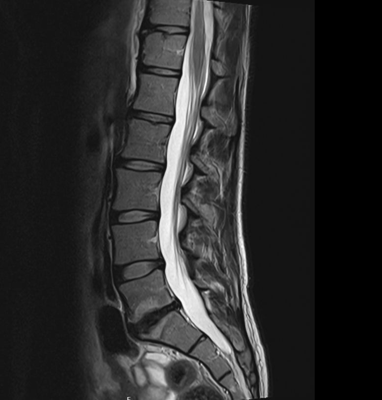 Show labels Enlarge
Sagittal lumbar MRI. Prominent Modic Type 2 endplate changes centered at the L5–S1 motion segment — involving the inferior endplate of L5 and the superior endplate of S1 — with associated degenerative disc disease, interpreted together with the clinical picture to support vertebrogenic pain. De-identified · no PHI.
- Treated vertebral levels
- L5 · S1
- #Imaging findings
- 1Modic Type 2 endplate changeInferior L5 endplate
- 2Modic Type 2 endplate changeSuperior S1 endplate
Reading the Imaging
The MRI is interpreted together with the patient's history, examination, and treatment response.
- Patient Overview
A 32-year-old woman — an active mother of two and former collegiate athlete — presented with persistent chronic axial low back pain that progressively limited the physically active lifestyle she had maintained since childhood. Before the onset of chronic vertebrogenic pain, she regularly participated in cycling, gym-based exercise, outdoor recreation, and other physically demanding activities that were central to her overall health and quality of life.
Despite prolonged conservative treatment — including physical therapy, a home exercise program, oral medications, anti-inflammatory medications, epidural steroid injections, facet joint injections, trigger point injections, chiropractic care, acupuncture, and activity modification — chronic axial low back pain continued to significantly impact function and her ability to remain physically active.
- Clinical Evaluation
Following comprehensive review of the patient's history, physical examination, characteristic pain pattern, and lumbar MRI, vertebrogenic pain was considered a significant contributor to her chronic axial low back pain.
Lumbar MRI demonstrated prominent Modic Type 2 endplate changes centered at the L5–S1 motion segment, involving the inferior endplate of L5 and the superior endplate of S1, with associated degenerative disc disease.
When interpreted together with the patient's history, physical examination, characteristic pain pattern, and prolonged unsuccessful response to conservative treatment, these findings supported vertebrogenic pain involving the L5 and S1 vertebral levels. A two-level Basivertebral Nerve Ablation was subsequently performed utilizing the Boston Scientific Intracept® system.
Fluoroscopic Images of Radiofrequency Ablation Probe Placement
Under fluoroscopic guidance, paired anteroposterior (AP) and lateral images were obtained to localize each level and to confirm final radiofrequency ablation (heater) probe position within the vertebral body — S1 and L5 — using the Boston Scientific Intracept System.

Anteroposterior (AP) view
S1 vertebral body
Anteroposterior (AP) image demonstrating fluoroscopic localization of the S1 vertebral body.

Lateral view
S1 vertebral body
Lateral image confirming final positioning of the Boston Scientific Intracept heater probe within the S1 vertebral body before completion of treatment.

Anteroposterior (AP) view
L5 vertebral body
Anteroposterior (AP) image demonstrating fluoroscopic localization of the L5 vertebral body.

Lateral view
L5 vertebral body
Lateral image confirming final positioning of the Boston Scientific Intracept heater probe within the L5 vertebral body.
The Clinical Course
Improvement following BVNA is frequently progressive; early symptoms are not the final outcome.
- Early Recovery
At the one-week postoperative follow-up, the patient reported approximately 30% improvement in previously described axial low back pain, with steadily improving postoperative discomfort. Recovery progressed appropriately without procedure-related complications.
The patient was counseled that continued improvement commonly occurs during the weeks following Basivertebral Nerve Ablation.
- Progressive Recovery
At the four-week postoperative evaluation, the patient reported approximately 70% improvement in chronic axial low back pain, with substantially improved activity tolerance.
- Long-Term Outcome
By approximately two months following the procedure, the patient reported approximately 95% resolution of the chronic axial low back pain that had previously limited her daily activities. She described returning to cycling, gym-based exercise, outdoor recreational activities, and the active lifestyle that had always been an important part of her life.
She also reported an improved ability to care for her family while returning to the activities that had previously provided enjoyment, stress relief, and overall well-being.
At a Glance
≈95%
resolution of chronic axial low back pain by ~2 months
Patient
32-year-old female · active mother of two, former collegiate athlete
Diagnosis
Chronic vertebrogenic low back pain
Prior treatment
Extensive unsuccessful conservative care
Procedure
Two-level BVNA (L5, S1), Boston Scientific Intracept® system
One week / four weeks
≈30% → ≈70% improvement
Two months
≈95% improvement · returned to cycling and exercise
Clinical Pearl
For many physically active patients, successful treatment of vertebrogenic pain is measured by restoration of function rather than pain scores alone. Returning to meaningful physical activity often represents one of the greatest improvements in quality of life following successful Basivertebral Nerve Ablation. Careful patient selection, appropriate MRI correlation, and comprehensive clinical evaluation remain fundamental to achieving favorable outcomes.
Continue Your Learning
Continue exploring the educational library at your own pace.
Have similar symptoms?
Every case is individual. A personal evaluation can help determine whether an assessment for vertebrogenic pain may be appropriate for you.