Returning to Community Life Through Treatment of Multilevel Vertebrogenic Pain
Clinical Question
Can careful clinical evaluation identify multilevel vertebrogenic pain in a patient with persistent chronic axial low back pain despite extensive conservative treatment and multilevel degenerative spinal disease?
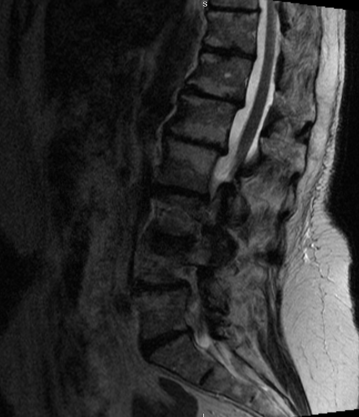
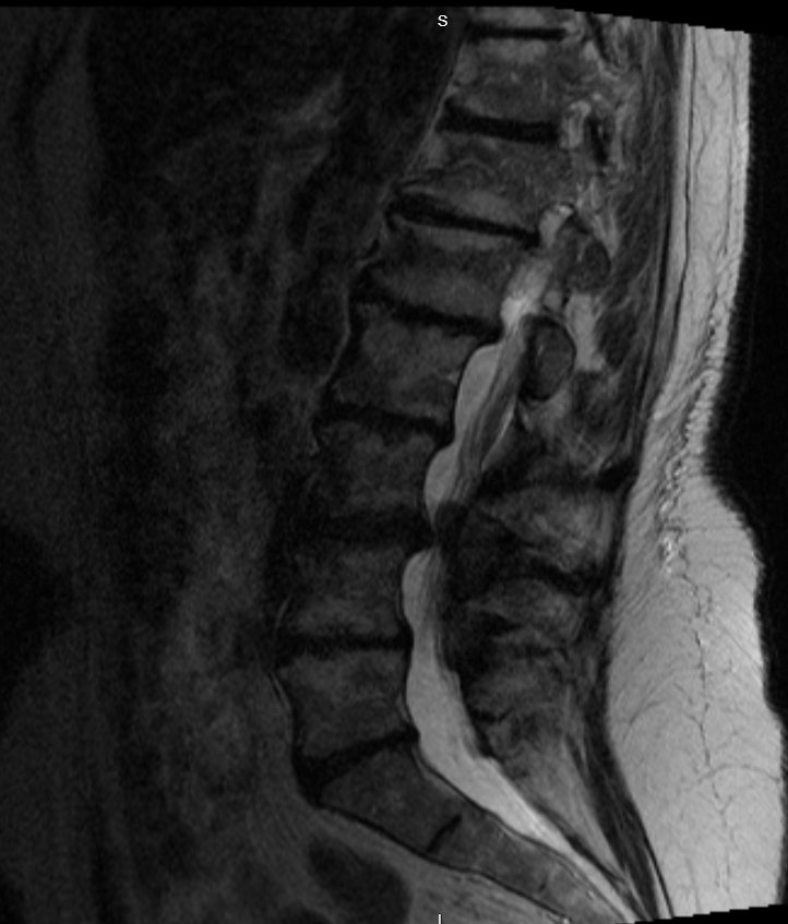
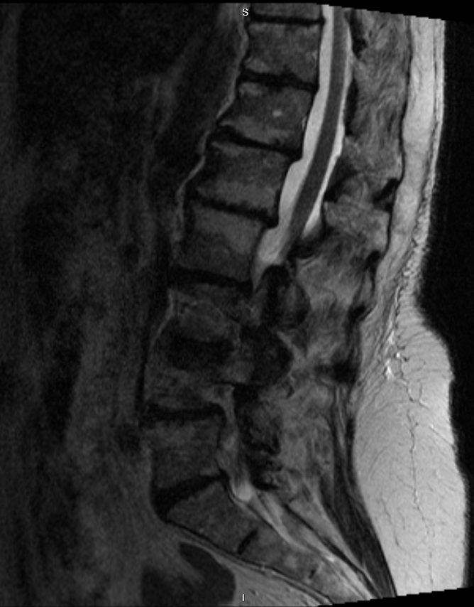
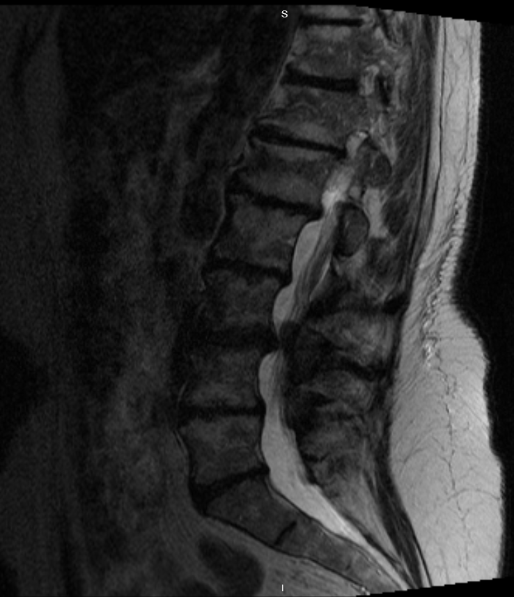 Show labels Enlarge
Sagittal lumbar MRI. Multilevel Modic Type 2 endplate changes extending from T12 through S1, with multilevel degenerative disc disease and disc height loss, interpreted together with the clinical picture to support multilevel vertebrogenic pain. De-identified · no PHI.
- Treated vertebral levels
- T12 · L1 · L2 · L3 · L4 · L5 · S1
- #Imaging findings
- 1Modic Type 2 endplate changesUpper levels — T12, L1, L2
- 2Modic Type 2 endplate changesMid levels — L3, L4
- 3Modic Type 2 endplate changesLower levels — L5, S1
Reading the Imaging
The MRI is interpreted together with the patient's history, examination, and treatment response.
- Patient Overview
A 78-year-old woman presented with persistent chronic axial low back pain that had progressively limited her daily activities and quality of life despite extensive conservative treatment.
Previous management included physical therapy, a home exercise program, oral medications, anti-inflammatory medications, oral corticosteroids, epidural steroid injections, facet joint injections, and activity modification. Despite these treatments, she continued to experience function-limiting axial low back pain affecting walking, prolonged standing, movement, bending, and routine daily activities.
- Clinical Evaluation
Comprehensive review of the patient's history, physical examination, characteristic pain pattern, and lumbar MRI demonstrated findings consistent with chronic vertebrogenic pain.
Lumbar MRI demonstrated multilevel Modic Type 2 endplate changes extending from T12 through S1, lumbar levoscoliosis centered at L3–L4, multilevel degenerative disc disease, multilevel disc height loss, chronic vertebral endplate remodeling, multilevel facet arthropathy, and multilevel foraminal stenosis.
The Modic endplate changes, interpreted together with the patient's history, physical examination, characteristic pain pattern, and unsuccessful response to prolonged conservative treatment, supported multilevel vertebrogenic pain. A multilevel Basivertebral Nerve Ablation was subsequently performed across the affected vertebral levels, from T12 through S1, utilizing the Stryker OptaBlate® system.
Fluoroscopic Images of Radiofrequency Ablation Probe Placement
Under fluoroscopic guidance, anteroposterior (AP) and lateral images were obtained to localize each level and to confirm radiofrequency ablation (heater) probe position within the vertebral body, treating from the sacrum upward through the low thoracic spine — S1 through T12 — using the Stryker OptaBlate® system.

Anteroposterior (AP) view
L5 and S1 vertebral bodies
Anteroposterior (AP) image demonstrating fluoroscopic localization and positioning of the Stryker OptaBlate heater probes within the L5 and S1 vertebral bodies.

Lateral view
L5 and S1 vertebral bodies
Lateral image of the same L5 and S1 levels, confirming the depth of the Stryker OptaBlate heater probes within the vertebral bodies.

Anteroposterior (AP) view
S1 and L4 vertebral bodies
Anteroposterior (AP) image demonstrating the Stryker OptaBlate heater probes positioned within the S1 and L4 vertebral bodies.

Anteroposterior (AP) view
L3 vertebral body
Anteroposterior (AP) image demonstrating the Stryker OptaBlate heater probe positioned within the L3 vertebral body.

Anteroposterior (AP) view
L1 and L2 vertebral bodies
Anteroposterior (AP) image demonstrating the Stryker OptaBlate heater probes positioned within the L1 and L2 vertebral bodies.

Lateral view
L1 and L2 vertebral bodies
Lateral image of the same L1 and L2 levels, confirming the depth of the Stryker OptaBlate heater probes within the vertebral bodies.

Lateral view
T12 and L3 vertebral bodies
Lateral image confirming the depth of the Stryker OptaBlate heater probes within the T12 and L3 vertebral bodies.

Anteroposterior (AP) view
T12 vertebral body
Anteroposterior (AP) image demonstrating final positioning of the Stryker OptaBlate heater probe within the T12 vertebral body.
The Clinical Course
Improvement following BVNA is frequently progressive; early symptoms are not the final outcome.
- Early Recovery
At the one-week postoperative follow-up, the patient reported approximately 20% improvement in her previously described axial low back pain. Recovery was appropriate, and no procedure-related complications were identified.
She was counseled that improvement following Basivertebral Nerve Ablation is frequently progressive over the weeks following treatment.
- Progressive Recovery
At the one-month follow-up, the patient reported approximately 40% improvement in her chronic axial low back pain, with steadily improving activity tolerance.
- Long-Term Outcome
By approximately two months following the procedure, the patient reported an estimated 60% reduction in the chronic axial low back pain that had previously limited her quality of life. She described meaningful improvement in standing tolerance, walking tolerance, and routine daily activities.
Most importantly, she was able to resume community volunteer activities that had become increasingly difficult because of her chronic vertebrogenic pain, including volunteering with local animal rescue organizations and participating in community service activities that had previously brought significant purpose and enjoyment to her life. She described these improvements as restoring an important part of her independence and quality of life.
At a Glance
≈60%
reduction in chronic axial low back pain by ~2 months
Patient
78-year-old female
Diagnosis
Chronic vertebrogenic low back pain
Prior treatment
Extensive unsuccessful conservative care
Procedure
Multilevel BVNA (T12–S1), Stryker OptaBlate® system
One week / one month
≈20% → ≈40% improvement
Two months
≈60% improvement · returned to community volunteer work
Clinical Pearl
Patients often describe successful treatment in terms of the activities they regain rather than the amount of pain they lose. For many individuals with vertebrogenic pain, meaningful improvement is measured by returning to family responsibilities, hobbies, volunteer work, and community participation. Careful patient selection remains fundamental to identifying those most likely to benefit from Basivertebral Nerve Ablation.
Continue Your Learning
Continue exploring the educational library at your own pace.
Have similar symptoms?
Every case is individual. A personal evaluation can help determine whether an assessment for vertebrogenic pain may be appropriate for you.