See What Causes Vertebrogenic Pain
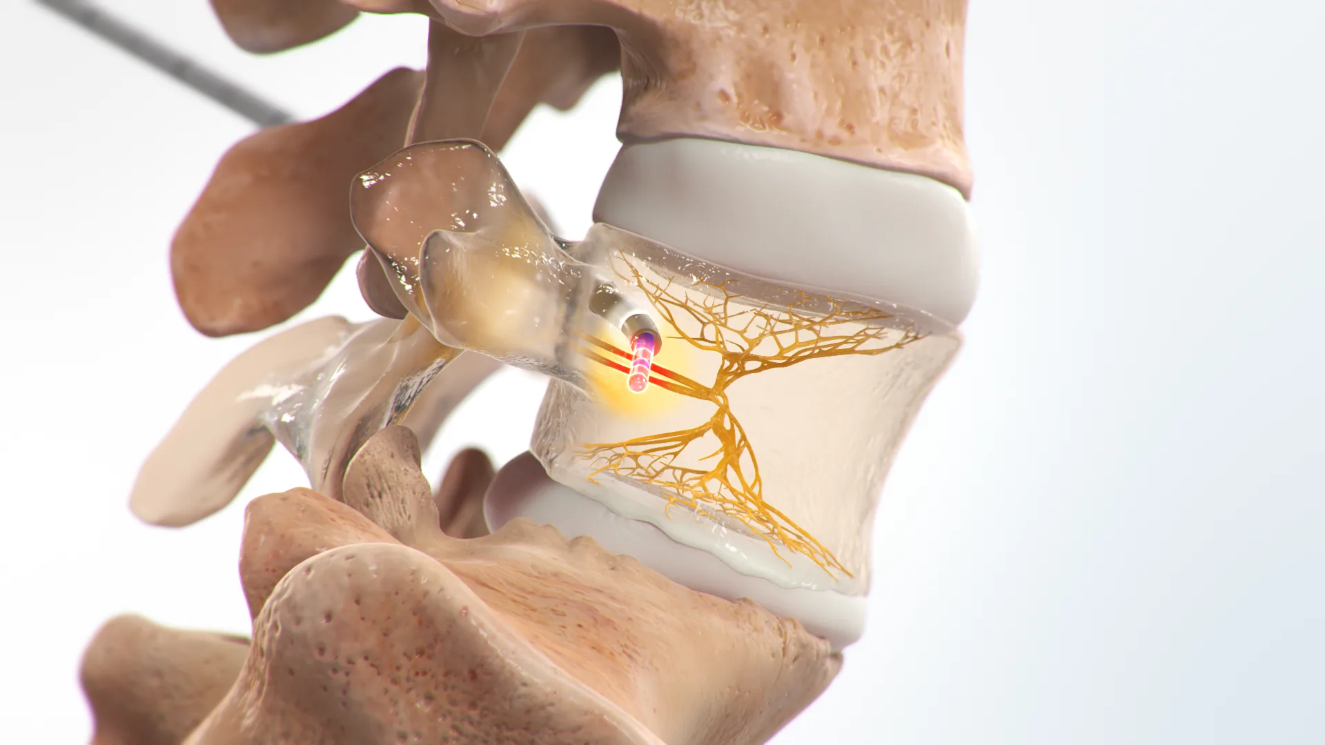
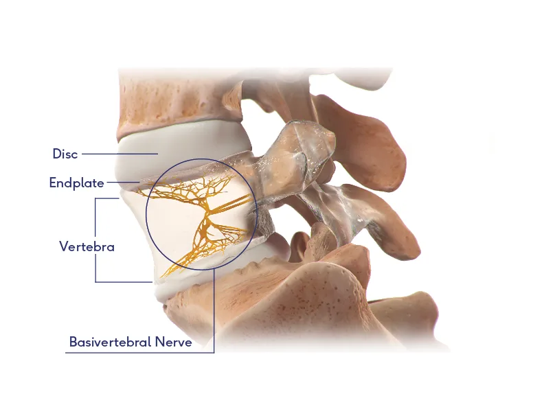
Vertebrogenic pain comes from the vertebra itself — specifically the endplates and the basivertebral nerve inside the bone. Explore the anatomy below, then step through how this kind of pain is thought to develop.
What You'll Learn
Approx. 4–6 min read- The key structures: disc, endplate, vertebra, and basivertebral nerve
- How everyday wear can lead to vertebrogenic pain, step by step
- Where these changes can appear on an MRI
- How this connects to the BVNA procedure
The Anatomy of a Spinal Segment
Select a point to learn what each structure does and why it matters in vertebrogenic pain.
How Vertebrogenic Pain Develops
Follow the vertebral endplate from a healthy spine to the moment the basivertebral nerve carries a pain signal. Use the steps, arrows, or arrow keys to move through the sequence.
- 1Healthy disc
- 2Degeneration
- 3Endplate damage
- 4Inflammation
- 5Pain signal
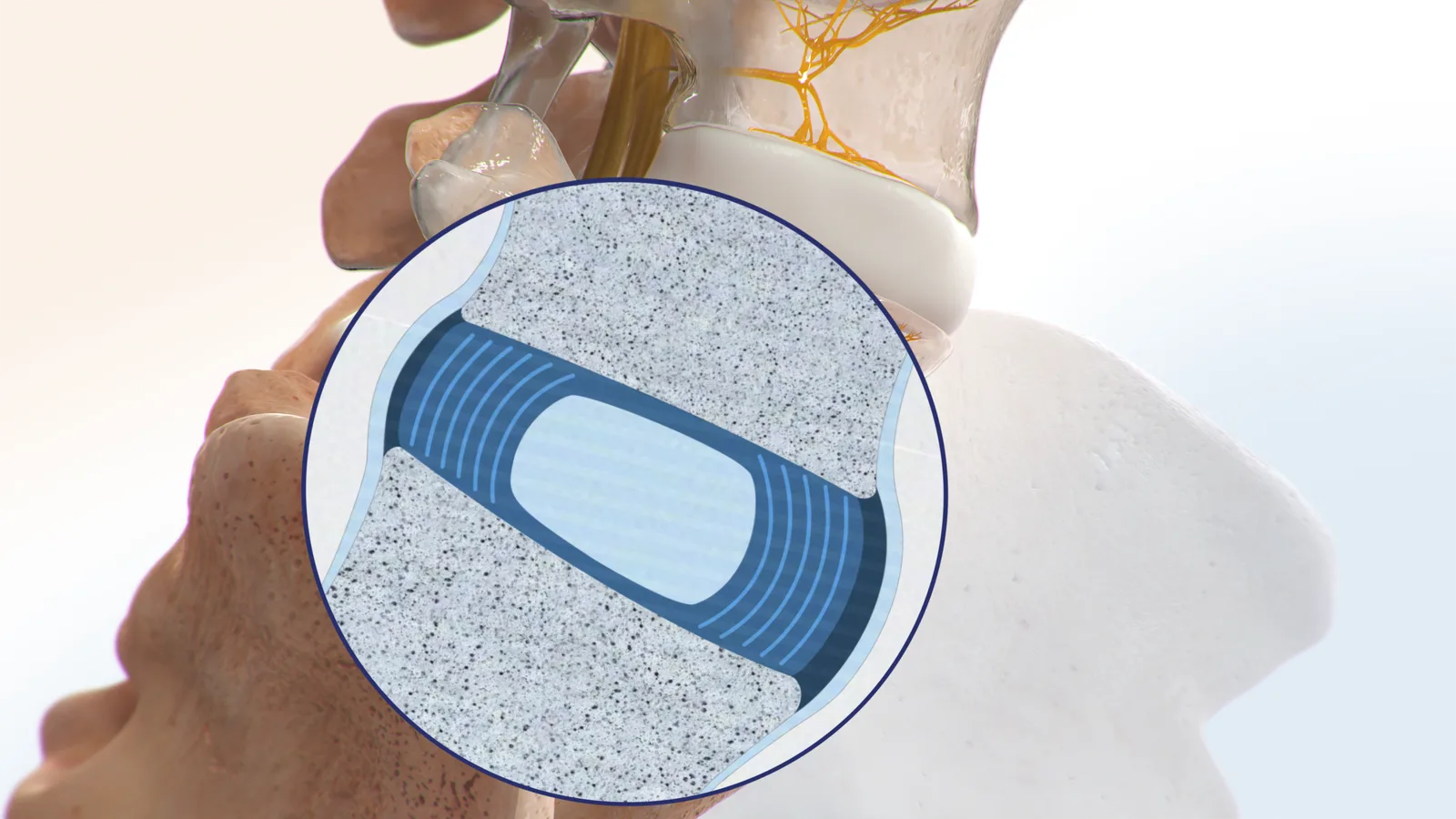
A healthy disc cushions the spine
Between each pair of vertebrae sits an intervertebral disc that acts as a shock absorber. A healthy disc is well hydrated, and the vertebral endplates — the thin layers of tissue covering the top and bottom of each vertebral body — are smooth and intact.
This image sequence follows the endplate up close. For an animated, scroll-driven look at how the discs themselves change over time, see Could My Pain Be Vertebrogenic?
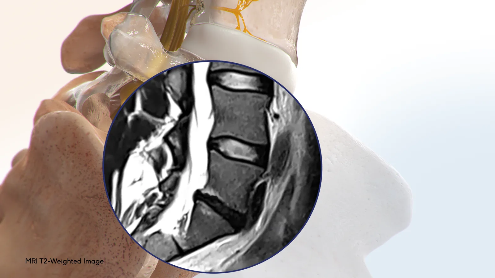
Where It Shows Up on an MRI
The same vertebral endplates can be examined on an MRI. Changes near the endplates — called Modic changes — are one clue clinicians may weigh alongside your history and examination.
- MRI looks at the vertebral endplates, where vertebrogenic changes occur.
- Modic Type 1 and Type 2 are two endplate patterns clinicians may look for.
- Imaging is one piece of the picture — never a diagnosis on its own.
Four Structures, One Pain Pathway
Vertebrogenic pain is best understood as a chain: wear on the disc stresses the endplates, the endplates become inflamed, and the basivertebral nerve carries that signal.
Disc
The shock absorber that wears down with everyday living.
Endplate
The tissue layer that becomes damaged and inflamed.
MRI clue
Modic changes that may be visible near the endplates.
Basivertebral nerve
The pathway that carries the pain — and the BVNA target.
Continue Your Learning
Keep exploring the educational library at your own pace.
Ready to Understand Your Own Pain?
If this sounds like your experience, an evaluation can help determine what may be contributing to your chronic low back pain.