MRI Learning Center
How to read a lumbar spine MRI and recognize Modic endplate changes — the imaging clue that axial low back pain may be coming from a specific, treatable source at the vertebral endplates.
What You'll Learn
Approx. 8–10 min read- How to tell a T1 image from a T2 image at a glance
- How to find the vertebral endplates on a sagittal MRI
- How to recognize Modic Type 1, 2, and 3 changes
- Why T1 or STIR — not T2 alone — separates Type 1 from Type 2
- How endplate changes connect to vertebrogenic pain and BVNA
Reading a Real MRI
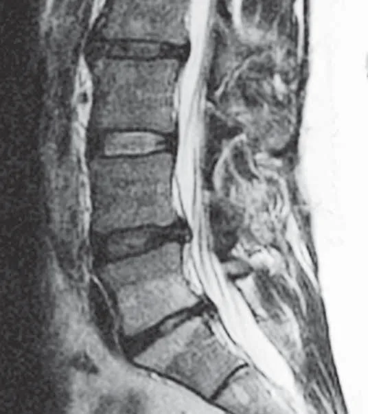
This is a de-identified sagittal T2-weighted lumbar MRI demonstrating Modic endplate changes at the L4–L5 level. Follow the same four steps a radiologist uses to read it.
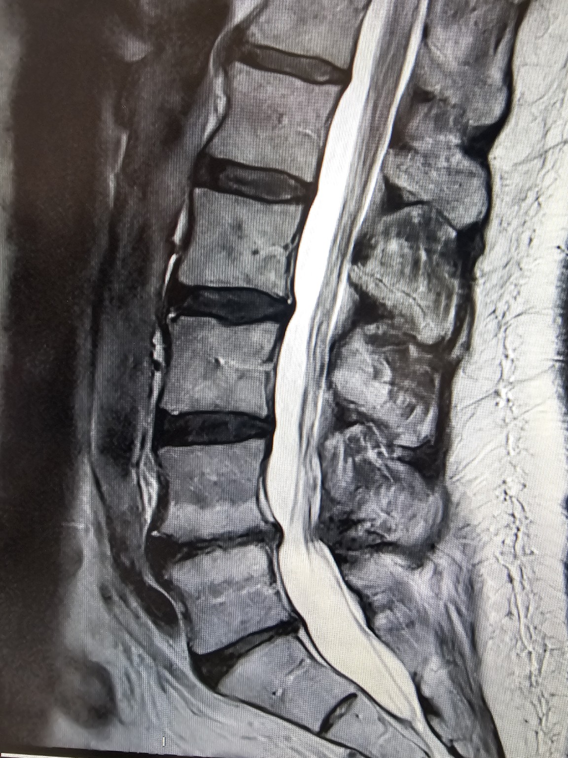
What you are seeing
The vertebral bodies are stacked in profile with the discs between them, and the bright vertical stripe of cerebrospinal fluid (CSF) runs down the spinal canal. Because that fluid is bright, this is a T2-weighted image.
At the L4–L5 level, the bone marrow immediately beneath the endplates shows altered signal — the hallmark location of a Modic change, part of the degenerative process at that motion segment.
Note the discipline of the read: identify the plane, confirm the weighting from the CSF, locate the endplates, then look for signal hugging them. The steps below generalize this to any lumbar MRI.
How to Read a Spine MRI, Step by Step
Four steps take you from a screen full of grey to a confident read of the endplates.
1 · Recognize the plane
A sagittal image is the 'spine from the side' view — vertebral bodies stacked front-to-back, discs between them, and the spinal canal behind. (Axial = cross-sections; coronal = front-facing.)
2 · Decide T1 or T2
Look at the CSF in the canal. Bright CSF = T2 ('water-weighted'). Dark CSF with bright body fat = T1 ('fat-weighted'). This single check anchors everything that follows.
3 · Find the endplates
The endplates are the flat top and bottom surfaces of each vertebral body where it meets the disc. Normal marrow sits just inside them; that subchondral zone is where Modic changes live.
4 · Spot signal hugging the endplate
Look for a band of abnormal signal in the marrow right against the endplate, usually at a degenerated disc level — then classify it using T1 + T2 (and STIR) as shown below.
The Three Modic Types on MRI
Modic changes are graded by how the subchondral marrow behaves across MRI sequences. 'Hyperintense' means bright; 'hypointense' means dark. A useful memory aid: Type 1 is dark-then-bright, Type 2 is bright-then-bright (it's fat), Type 3 is dark-then-dark.
| Sequence | Type 1 — edema / inflammatory | Type 2 — fatty marrow | Type 3 — sclerotic |
|---|---|---|---|
| T1-weighted | Hypointense (dark) | Hyperintense (bright) | Hypointense (dark) |
| T2-weighted | Hyperintense (bright) | Iso– to mildly hyperintense | Hypointense (dark) |
| STIR (fat-suppressed) | Hyperintense (stays bright) | Hypointense (suppressed) | Hypointense (dark) |
| Underlying tissue | Fibrovascular edema / inflammation (active) | Yellow fatty marrow replacement (more chronic) | Dense subchondral bone sclerosis |
Why T2 Alone Cannot Separate Type 1 from Type 2
On T2, both edema and fat look bright.
A bright endplate lesion on a T2 image is ambiguous — it could be inflammatory edema (Type 1) or fatty marrow (Type 2). To tell them apart you need another sequence:
- T1 separates them. Type 1 is dark on T1 (edema); Type 2 is bright on T1 (fat). This is the single most useful discriminator.
- STIR separates them. STIR nulls fat, so Type 2 (fatty) goes dark while Type 1 (edema) stays bright.
Rule of thumb: a T2-bright endplate lesion that suppresses on STIR is fat (Type 2); one that stays bright on STIR is edema (Type 1).
Type 1 and Type 2 on MRI
MRI scans are read in two common “weightings” — called T1 and T2 — that make tissues look lighter or darker in different ways. Radiologists describe Modic changes by the pattern they show across both. Neither type is “better” or “worse”; they are simply different appearances a clinician interprets in context.
Modic Type 1
Often a more “active” appearance
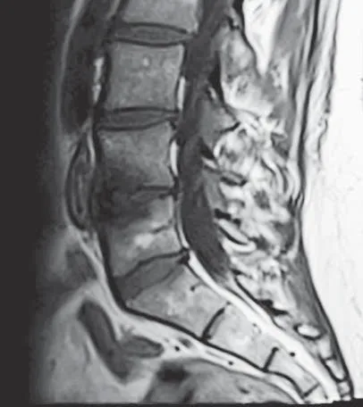
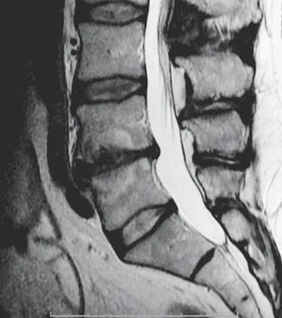
On a T1-weighted image the endplate change tends to look darker; on a T2-weighted image it tends to look brighter.
Modic Type 2
Often a more “settled” appearance
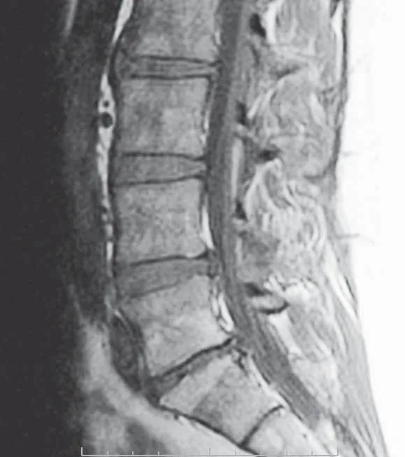
On both T1- and T2-weighted images the endplate change tends to look brighter.
De-identified MRI examples © Boston Scientific — physician-authorized educational use.
From Endplate Signal to a Treatable Pain Source
Damaged vertebral endplates can become a distinct source of chronic axial (midline) low back pain. Pain signals from the endplates travel through the basivertebral nerve (BVN) — the target of basivertebral nerve ablation (BVNA).
The basivertebral nerve
The BVN enters the back of the vertebral body and runs toward its center, then branches toward the superior and inferior endplates. When endplates are damaged, inflammation and nerve ingrowth can make them a source of pain — and Modic changes are the MRI signature of that process.
How candidates are identified
Published selection criteria pair imaging with the clinical picture: chronic axial low back pain for at least six months, unresponsive to at least six months of conservative care, with Modic Type 1 or Type 2 changes on MRI (typically L3–S1). Modic Type 3 is not a target.
Look Beyond the Midline
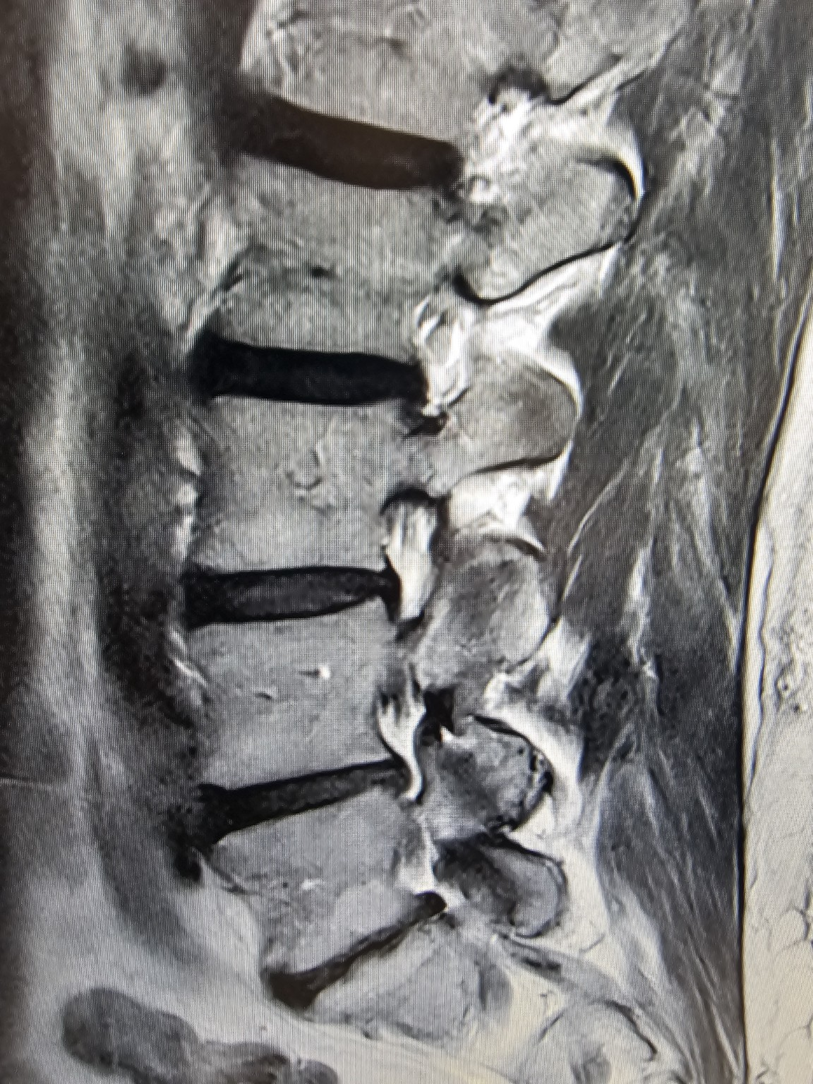
Sagittal MRI is a stack of slices from one side to the other. Endplate changes are not always symmetric — sometimes the most significant Modic changes sit off to one side, and appear only on the more lateral (parasagittal) slices.
The great masquerader
This patient had pain radiating to the right side, in a pattern that looked clinically like SI joint dysfunction, radiculopathy, or sciatica. The true source was vertebrogenic — degenerative disc disease with Modic endplate changes activating the basivertebral nerve — but the significant changes were concentrated on the right side of the vertebral body.
On the midline and left-sided slices, the endplate changes look subtle. Only when you scroll across the sagittal images from right to left do the significant Modic changes come into view — which is exactly why this more lateral slice is the informative one.
Two takeaways
- Review the whole stack. Don't judge the endplates from the midline slice alone — scroll laterally in both directions.
- Correlate with the exam. Vertebrogenic pain can convincingly mimic other diagnoses; imaging and the clinical picture have to be read together.
Where the Change Sits — and Its Shape — Matter
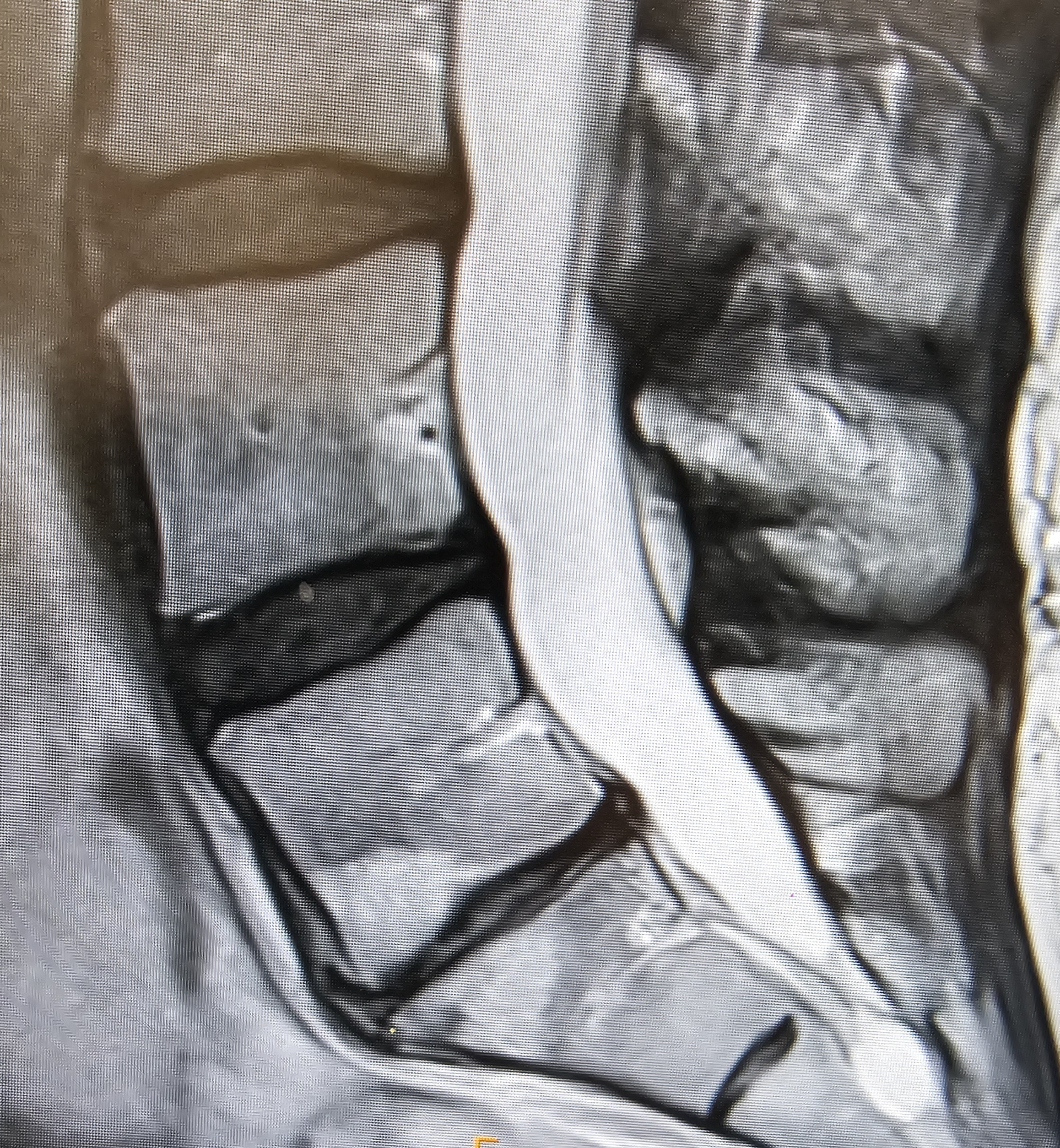
Modic changes are not only graded by type; their location and shape carry information too. This close-up of a single disc level shows how the pattern can point back to a patient's specific symptoms.
Shape follows load
Here the endplate changes form a wedge that is widest at the front of the vertebral bodies. That anterior concentration mirrors where the spine is loaded most during certain movements.
This patient reported that low back pain increased with forward flexion — bending forward shifts load onto the anterior column, exactly where these changes are most pronounced. The morphology and the symptom pattern tell a consistent story.
What to take from it
- Note the location. Anterior, posterior, or across the whole endplate — where the change sits can align with how the pain behaves.
- This is single-segment disease. The Modic changes here involve one disc level — the L5 and S1 endplates around the L5–S1 disc.
Imaging Is One Part of the Picture
Modic changes are common and are frequently found in people without pain — so their presence does not, by itself, prove they are the source of a person's symptoms.
MRI findings are only meaningful when correlated with the history, physical examination, pain pattern, and response to prior treatment. Early spinal infection can also mimic Type 1 changes and must be excluded when the clinical picture warrants.
This page is educational and is not a substitute for evaluation by a qualified clinician.
Terms Worth Knowing
Endplate
The flat top and bottom 'caps' of each vertebral body, where bone meets the cushioning disc.
Subchondral
The layer of bone just beneath a joint surface. Modic changes live in the subchondral bone under the endplate.
Sagittal
A side-on, vertical view that slices the body into left and right — the 'spine from the side' picture.
T1-weighted
An MRI setting where fat looks bright and fluid looks dark. Great for anatomy; CSF is dark.
T2-weighted
An MRI setting where fluid looks bright. Good for showing swelling and disc problems; CSF is bright.
STIR
A fat-suppressing MRI setting that turns fat dark so swelling (edema) lights up and is easy to see.
Hyperintense / Hypointense
Hyperintense means it appears bright; hypointense means it appears dark.
Edema
Fluid or swelling in tissue — a sign of active inflammation. Bright on T2 and STIR.
Frequently Asked Questions
Educational answers to a few common questions about reading an MRI for Modic changes.
Can a T2 image alone tell whether a change is Modic Type 1 or Type 2?
Do Modic changes always cause back pain?
Which Modic type is most associated with pain?
Where do Modic changes usually appear?
Does finding Modic changes mean I need a procedure?
References & Further Reading
Selected peer-reviewed and radiology references used to compile this page. Educational use only.
- [1]Modic MT, et al.. Degenerative disk disease: assessment of changes in vertebral body marrow with MR imaging. Radiology (classification origin) — reviewed in Radsource: Vertebral Endplate (Modic) Changes. 1988 / ref..Link
- [2]Review. Modic changes — pathobiology and clinical significance. PMC (peer-reviewed review). 2021.Link
- [3]Radiology Masterclass. MRI: understanding T1- and T2-weighted images. radiologymasterclass.co.uk. ref..Link
- [4]Review. STIR and fat suppression — image characterization. PMC / MRI reference. ref..Link
- [5]Lorio M, et al. (review). Vertebrogenic pain: the basivertebral nerve and endplate origin of chronic low back pain. Pain Medicine (Oxford). 2022.Link
- [6]StatPearls. Basivertebral Nerve Ablation. NCBI Bookshelf. current.Link
- [7]Fischgrund JS, et al. (SMART trial). Intraosseous basivertebral nerve ablation for chronic low back pain: randomized, double-blind, sham-controlled trial. European Spine Journal. 2018.Link
- [8]Khalil JG, Smuck M, et al. (INTRACEPT trial). A prospective, randomized, controlled trial of basivertebral nerve ablation vs. standard care. The Spine Journal. 2019.Link
Continue Your Learning
Keep exploring the educational library at your own pace.
Understanding your MRI is one step toward understanding your pain.
If chronic axial low back pain has persisted despite conservative care, a personal evaluation can help determine whether vertebrogenic pain may be a contributor.